Medical & Life Science
Laser beams in the medical field span a broad array of exciting established and new applications - laser eye surgery, nanoparticle irradiation for skin cancer treatment, and laser ablation to create scar tissue or remove kidney stones, tumors or growths.
In medical treatments, the laser must irradiate the targeted material at a specific wavelength for a specific intention, without harm to surrounding tissue. This means a focus that is accurate, with proper intensity distribution, and within procedural specifications.
Continuous or frequent laser beam monitoring is recommended to verify proper beam performance. For example, verifying the laser focus hasn’t shifted to an unintentional location, and that it is properly delivering power over the expected area and laser beam shape.
Regular beam profiling is recommended to verify key parameters. Information on laser beam parameters below.
Medical & Life Science – Laser Use Examples
Categorized by Electromagnetic Spectrum Region
UV | ✓ Ablation of Tissues, Eye Surgery ✓ LASIK ablation with pulsed excimer laser (~10 - 50 W) |
Visible | ✓ Fluorescence, Ophthalmology ✓ Retinal Photocoagulation with continuous wave green argon laser (~50 - 2000 mW) |
Near Infrared (NIR) & Infrared (IR) | ✓ Microscopy, Soft Tissue, Dermatology, Scar Tissue Removal ✓ Microscopy imaging with Ti:Sapphire (~0.1 - 3 W avg) ✓ Cut Tissue with Er:YAG pulsed (~5 - 30 W) |
Medical & Life Science – Important Beam Parameters
Intensity
Irradiance Fluence Continuous & Pulsed |
Adequate beam intensity distribution means sufficient energy transfer to the material for proper tissue treatment or detection. The wrong amount of irradiance or fluence could result in suboptimal treatments such as improper ablation, or in general, energy applied too broadly or narrowly. These outcomes should motivate regular laser beam profiling. The 2D beam profile to the left shows a Gaussian beam intensity profile where the intensity is greatest at the center, white, and decreases moving outward toward the outer circumference, blue. Profiles like this give relative information about the intensity distribution. For other beam shape profiles used in medical and life science processes, see beam shape, below. The power applied at the beam waist divided by the spot size also gives information about the power intensity (for continuous beams). Common laser power levels used in medical and life science industries vary greatly considering the many laser processes: ~mW – W+. Note — For a continuous beam, the terms intensity, irradiance or power density are used: power divided by area, W/cm². For a pulsed beam, the term fluence or energy density is used: energy divided by area, J/cm². A pulse, repeated at the pulse frequency, will have peak irradiance and maximum pulse energy values reached during the pulse. |
Beam Waist Spot Size Focus |
At the focus, the beam diameter reaches a minimum, often referred to as the spot size or beam waist diameter. Focusing a beam to a smaller spot size will increase the density in that spot and vice versa. It is important to apply the optimal amount of power or energy at the specified spot size. Too large or too small will affect the desired target location and may lead to some of the defects mentioned previously. Common spot size values for the industry vary from ~0.2 µm – cm² depending on the application. For example, skin treatments may use a large spot size. |
Focal Plane Focal Distance |
The focal plane of a non-collimated laser beam is generally where the beam is focused to its smallest spot size. The focal distance is the distance from the focusing lens along the axis of propagation to the focal plane and can vary depending on the presence of other optics: the laser source, focusing optics and possible beam shaping devices. The focal plane, in many cases, lines up exactly with the material surface or working plane, but may be offset, examples below:
|
Beam Shape
|
Common shapes:
Beam profilers provide a quick and effective means to quantify the relative intensity distribution of a beam to verify beam shape. |
Beam Propagation |
Beam propagation is the behavior of a laser beam propagating through free space and is described by M² (beam quality), divergence and pointing. M² characterizes how close a power intensity profile is to a “Gaussian” beam and can give a sense of how focused the beam is. Laser cutting is an example where a more focused beam is important, so lower M² values are desirable.
Divergence describes the angle the beam diverges outward from the beam waist into the far field, much beyond the Rayleigh length. In contrast, divergence near zero is a way to confirm a beam is collimated, for example before being focused. This helps to ensure that once the beam is focused, it will be at the correct spot size and location. Pointing is the angle of laser beam propagation with respect to the optical axis. A pointing value of zero means it is perfectly aligned with the optical axis. It characterizes how much a laser stays on center as it gets farther from the laser source, including accuracy and precision. Pointing measurements support better beam alignment. A misaligned laser can disrupt how much power is delivered to the target tissue or material, compromising medical and life science outcomes. Misalignments can be caused by thermal fluctuations in the environment or in the laser system of high power lasers, as well as due to attenuation and time. |
Have questions or need help identifying the right solution for your application?

Related Products
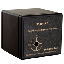
Beam'R2
- Affordable single-plane beam profiling
- Directly profile beams as small as 2 µm
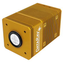
WinCamD-QD
- Options for 1550 and 2000 nm
- Large sensor formats available
WinCamD-LCM
- Versatile global shutter supports CW & pulsed beams
- 355 to 1150 nm standard with UV, 1310 nm, and 1550 nm options

BladeCam2-XHR
- Compact, affordable beam profiling
- Ultra-small 3.2 µm pixels
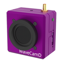
WaveCamD
- Shack-Hartmann wavefront sensor
- Zonal (numerical) and modal (Zernike) wavefront reconstruction